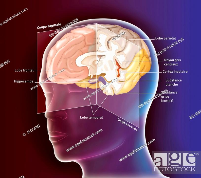
Stock Photo - Illustration of the anatomy of the brain, on a sagittal plane cross-section of the right hemisphere and a frontal cross-section of the left hemisphere, showing the hippocampus (purple), the insular cortex (orange), the frontal lobe (orange coloured), the parietal lobe (dark beige), the temporal lobe (yellow). AlzheimerÆs disease is caused by degeneration of the neurons and synapses in the cerebral cortex and some subcortical areas. This abnormal loss leads to atrophy of the affected areas, including the temporal and parietal lobes, part of the frontal cortex and the cingulate gyrus : the areas affecting speech and memory are affected.

Stock Photo: Illustration of the anatomy of the brain, on a sagittal plane cross-section of the right hemisphere and a frontal cross-section of the left hemisphere.

Searchable keywords

  • Alzheimer's Disease
  • Anatomical
  • Anatomy
  • Artwork
  • Basal
  • Basal Ganglia
  • Basales
  • Brain
  • Brain Cortex
  • Brain Hemisphere
  • Central Nervous System
  • Cephalic
  • Cerebral Cortex
  • Cerebral Hemisphere
  • Cerebrum
  • Claustrum
  • Cns
  • Cortex
  • Degenerate
  • Degeneration
  • Degenerative
  • Dementia
  • Drawing
  • Encephalon
  • Encephalus
  • Front
  • Frontal Lobe
  • Ganglion
  • Gray Matter
  • Grey Matter
  • Harmless
  • Harmlessly
  • Head
  • Healthy
  • Hemisphere
  • Hippocampus
  • Human
  • Illustration
  • Illustration of the anatomy of the brain
  • Insula
  • Insular
  • Limbic
  • Limbic System
  • Lobe
  • Mantle
  • Medicinal
  • Medicine
  • Nervous System
  • Neural
  • Neurological
  • Neurology
  • No Pathology
  • Normal
  • Nuclei
  • Nucleus
  • Outline
  • Pallium
  • Parietal
  • People
  • Rhinencephalon
  • Scheme
  • Senile Dementia
  • Sihouetted
  • Silhouette
  • Striatum
  • System
  • Telencephalon
  • Temporal Lobe
  • Ventral
  • White Matter
  • Without Pathology
Choose multiple keywords