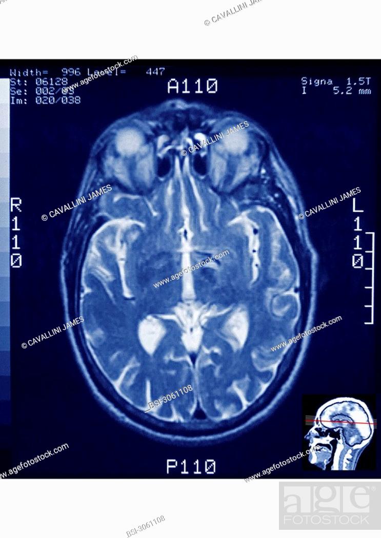
Stock Photo - Axial section of a human skull visualized by MRI _ Are visible : the brain, the eyes and the optic nerves, the sinuses.

Stock Photo: Axial section of a human skull visualized by MRI - Are visible : the brain, the eyes and the optic nerves, the sinuses.

Searchable keywords

  • Healthy
  • People
  • Human
  • Blue
  • Eye
  • Medicine
  • Examination
  • Examining
  • Head
  • Scan
  • Cell
  • Nuclear
  • Science
  • Result
  • Skull
  • Image
  • Brain
  • Examine
  • Investigate
  • Tobacco
  • Area
  • Mantle
  • Inspection
  • Medicinal
  • Socket
  • Investigation
  • Nuclear Magnetic Resonance
  • Normal
  • Posterior
  • Imaging
  • Technique
  • Eyeball
  • Lateralis
  • Orbit
  • Harmless
  • Lateral
  • Neurology
  • Imagery
  • Arterial
  • Artery
  • Vascular
  • Ventricle
  • Sem
  • Ethmoidal
  • Sinus
  • Cerebri
  • Neural
  • Cranial
  • Fissure
  • Anterior
  • Ocular
  • Neurological
  • Thalamus
  • Cerebrum
  • Encephalon
  • Cortex
  • Cortical
  • Ventricular
  • Oculi
  • Vascularization
  • Thalamic
  • Diencephalon
  • Meninges
  • Meninx
  • Encephalus
  • Cephalic
  • Arterio
  • Sulcus
  • Meningeal
  • Harmlessly
  • Arteria
  • Bulbus
  • Sylvius
  • Cns
  • Trigone
  • Habenulae
  • Ventriculus
  • Pallium
  • Betweenbrain
  • Diencephalic
  • Gray Matter
  • Grey Matter
  • Medical Examination
  • Nervous System
  • Nuclear Medicine
  • Temporal Lobe
  • Ethmoid Sinus
  • White Matter
  • Examination Technique
  • Blood Vessel
  • Investigation Technique
  • Magnetic Resonance
  • Magnetic Resonance Imaging
  • Medical Imaging
  • Medical Investigation Technique
  • Nasal Cavity
  • Nasal Fossa
  • Nasal Fossae
  • No Pathology
  • Occipital Lobe
  • Optic Nerve
  • Sylvian Fissure
  • Without Pathology
  • Brain Cortex
  • Central Nervous System
  • Cerebral Artery
  • Cerebral Cortex
  • Trigonum
  • Ventriculi
  • Habenular
Choose multiple keywords