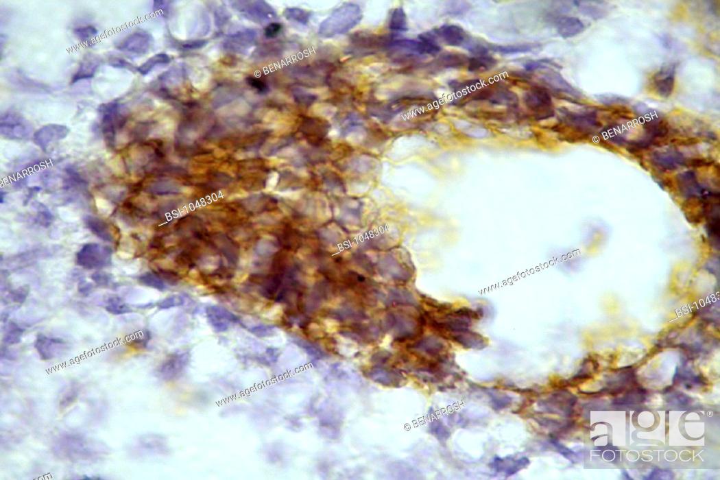
Photo de stock - FETAL LUNG, HISTOLOGY<BR>Histological section of a human fetal lung at the stage of 14 weeks of gestation. Staining with an anti cytokeratin 19 antibody, in brown, and with hematoxylin (for nuclear DNA), en blue. Magnification: 200x. At this stage the lung is immature and the future airways (e.g. bronchi) look like glands. The parenchyma and the forming cells (visualized by their nuclei in blue), have no significant density that usually is found in adults. The epithelium (colored in brown with an anti K19 (specific) antibody is in monolayer, not stratified and undifferentiated since there is no distinct cell types such as mucous, brush, etc. The cells within the parenchyma are developing. Some of the cells are in mallow – a colour resulting from the overlay of the nuclei which are in blue with the cytoplasm (at the sites of antibody signal).

Photo de stock: FETAL LUNG,  HISTOLOGY<BR>Histological section of a human fetal lung at the stage of 14 weeks of gestation.  Staining with an anti cytokeratin 19 antibody.

Mots-clés utilisés

Choisir des mots clés multiples