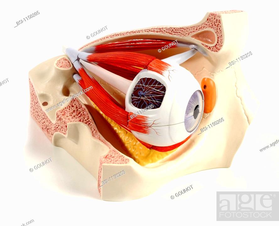
Stock Photo - Anatomic model of the human right eye three_quarter view. The eye, lying on the orbital fat in yellow, is enclosed in the orbit. This cavity is delimited mainly by the frontal orbital apex and side wall, sphenoid behind, zygomatic medial wall, t

Stock Photo: Anatomic model of the human right eye three-quarter view. The eye, lying on the orbital fat in yellow, is enclosed in the orbit.

Searchable keywords

  • Healthy
  • View
  • Sensitive
  • Eye
  • Medicine
  • Ophthalmology
  • Backgrounds
  • White
  • Sight
  • Vision
  • Fibre
  • Optic
  • Data
  • Document
  • Information
  • Plastic
  • Common
  • Sense
  • Muscle
  • Model
  • Coat
  • Plain
  • Visual
  • Medicinal
  • Bone
  • Socket
  • Jaw
  • Passage
  • Pupil
  • Detached
  • Anatomy
  • System
  • Inform
  • Aware
  • Upper
  • Visibility
  • Normal
  • Musculus
  • Documentation
  • Iris
  • Seeing
  • Eyesight
  • Eyeball
  • Bony
  • Apparatus
  • Cornea
  • Perception
  • Sensitivity
  • Orbit
  • Harmless
  • Tract
  • Fibrous
  • Sensory
  • Myology
  • Tunica
  • Sac
  • Neural
  • Anatomical
  • Musculature
  • Tendon
  • Ligament
  • Osseous
  • Afferent
  • Ocular
  • Ophthalmic
  • Annular
  • Sinew
  • Anatomic
  • Extrinsic
  • Informative
  • Oculi
  • Corneal
  • Perceptive
  • Maxillary
  • Lachrymal
  • Maxilla
  • Sclera
  • Ophthalmological
  • Fibrilla
  • Sensorial
  • Detachable
  • Motility
  • Sphenoid
  • Lacrymal
  • Lacrimal
  • Oculomotor
  • Sclerotic
  • Extra-Ocular
  • Harmlessly
  • Scleral
  • Bulbus
  • Trochlea
  • Zinn
  • Motricity
  • Sclerotica
  • Sphenoidal
  • Nervous System
  • Peripheral Nervous System
  • Muscle Fiber
  • Muscle Fibre
  • Muscular System
  • Myofibrillae
  • No Pathology
  • Optic Nerve
  • Sphenoid Bone
  • Tunica Albuginea
  • Upper Jaw
  • Vision Correction
  • Visual Perception
  • Visual System
  • Without Pathology
  • Pns
  • Myofilament
  • Viewed
  • Nerve
  • Muscular
Choose multiple keywords